Yannick Becker, Olivier Coulon and Adrien Meguerditchian
Language is in the heart of what is generally accepted to make us special as a species. The “How did it evolve?” persists hereby as one of themost debated questions in science. Many authors have stated that the difference between humans and non-human primates might lay in a different neural architecture between primate species (Boe et al., 2017, Friederici,2017) including one of its key feature: its connectivity.
In the human brain, the classic language pathway model describes a fibre pathway – the Arcuate fasciculus (AF) – arching around the sylvian fissure, linking superior temporal language regions (roughly Wernicke’s area) to ventrolateral frontal regions (roughly Broca’sarea). Not surprisingly, it was observed that interrupting lesions of the AF leads to clinical symptoms. Traditionally, AF lesions are associated with conduction aphasia, characterised by the inability to repeat words while speaking.
This major language brain connection matures during childhood into a dominant and left-hemisphere lateralised pathway (Dubois et al., 2009; Perani et al., 2011). While the architecture for language brains regions is highly conserved between primate species, main morphological changes happened in the AF, explaining our uniqueness to process language (Rilling et al., 2008). Some authors extend this hypothesis to propose that the AF with parts of Broca’s area is the biological manifestation of Chomsky’s controversial “Universal Grammar” (Friederici et al. 2017; Zaccharella and Friederici, 2017).
In other words, the AF is at the heart of the debate about what makes us human.
In this review, we briefly and didactically present the historical findings and techniques that advanced knowledge about primate neuroanatomy and the Arcuate Fasciculus. We also present the different terminology used in this domain and highlight how conclusions about AF evolution depend on nomenclature. In a second time, we will review studies concerning the continuity or discontinuity between human and non-human primates. The debate is centred around three main controversies:
1. Inferior Frontal Terminations: An evolutionary continuity between primate species stipulates its locations in the monkeys’ Broca’s homologue, whereas a discontinuity stipulates its locations rather in the monkeys’ superior frontal areas but not in Broca’s homologue.
2. Extended temporal lobe terminations: An evolutionary continuity between primate species stipulates its locations deep in the monkey’s inferior and ventral temporal lobe whereas a discontinuity stipulates more posterior and dorsal termination in the monkeys’ parieto-temporal junction.
3. Lateralisation: An evolutionary continuity between primate species stipulates the left lateralisation of the monkeys’ AF whereas a discontinuity stipulates an absence or inverse lateralisation.
Keywords:
– language evolution
– white matter
– fiber tracts
– hemispheric specialization
References
Boë, Louis-Jean, Frédéric Berthommier, Thierry Legou, Guillaume Captier, Caralyn Kemp, Thomas R. Sawallis, Yannick Becker, Arnaud Rey, and Joël Fagot. 2017. ‘Evidence of a Vocalic Proto-System in the Baboon (Papio Papio) Suggests Pre-Hominin Speech Precursors’. PLOS ONE 12(1):e0169321. doi: 10.1371/journal.pone.0169321.
Dubois, J., L. Hertz-Pannier, A. Cachia, J. F. Mangin, D. Le Bihan, and G. Dehaene-Lambertz. 2009. ‘Structural Asymmetries in the Infant Language and Sensori-Motor Networks’. Cerebral Cortex 19(2):414–23. doi: 10.1093/cercor/bhn097.
Friederici, Angela D. 2017.Language in Our Brain: The Origins of a Uniquely Human Capacity. Cambridge, Massachusetts: The MIT Press.
Friederici, Angela D., Noam Chomsky, Robert C. Berwick, Andrea Moro, and Johan J. Bolhuis. 2017. ‘Language, Mind and Brain’. Nature Human Behaviour 1(10):713–22. doi: 10.1038/s41562-017-0184-4.
Perani, D., M. C. Saccuman, P. Scifo, A. Anwander, D. Spada, C. Baldoli, A. Poloniato, G. Lohmann, and A. D. Friederici. 2011. ‘Neural Language Networks at Birth’. Proceedings of the National Academy of Sciences 108(38):16056–61. doi: 10.1073/pnas.1102991108.
Rilling, James K., Matthew F. Glasser, Todd M. Preuss, Xiangyang Ma, Tiejun Zhao, Xiaoping Hu, and Timothy E. J. Behrens. 2008. ‘The Evolution of the Arcuate Fasciculus Revealed with Comparative DTI’. Nature Neuroscience 11(4):426–28. doi: 10.1038/nn2072.
Zaccarella, E., and A. D. Friederici. 2017. ‘The Neurobiological Nature of Syntactic Hierarchies’.Neuroscienceand Biobehavioral Reviews81(Pt B):205–12. doi: 10.1016/j.neubiorev.2016.07.038.
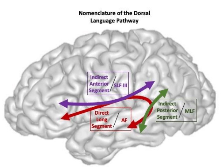
Figure 1: Schematic illustration of the dorsal language pathway terminology in the human brain. SLFIII: Third branche of the Superior Longitudinal Fasciculus, AF: Arcuate Fasciculus, MLF: Middle Longitudinal Fasciculus
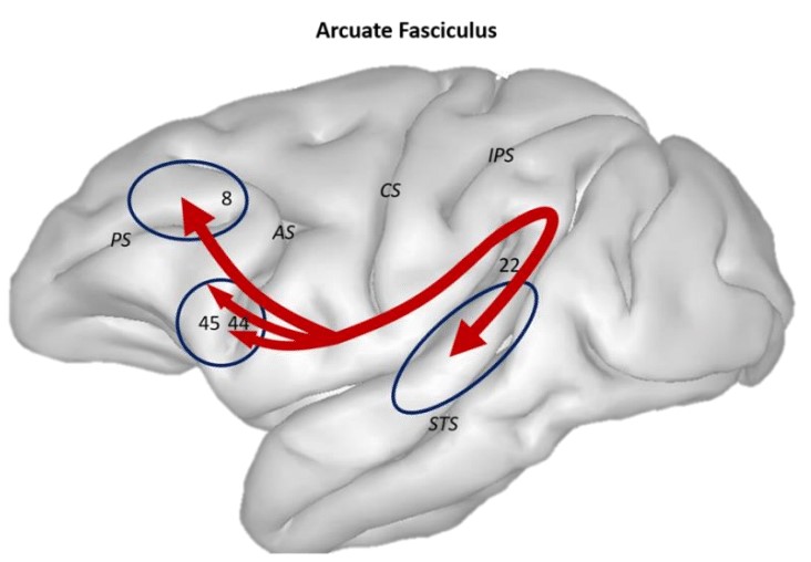
Figure 2: Illustration of thecourse andterminations (arrowheads) of the Arcuate Fasciculus (AF) in red. Blue circles are indicating thepossible terminations as discussed in the literature. PS: Principalis Sulcus, AS: Arcuate Sulcus, CS: Central Sulcus, IPS: Inferior Parietal Sulcus, STS: Superior Temporal Sulcus, Area 45 and 44, parts of Broca’s area, Area 22 part of Wernicke’s area.